Have you ever heard of flow cytometry? It's a technology that allows for the analysis of single...
Fluorescent Protein Cell Mimics as Positive Controls for Flow Cytometry Applications
.png?width=1200&height=400&name=Introducing%20HyParComp%20(6).png) Fluorescent proteins such as enhanced green fluorescent protein (eGFP) and monomeric Cherry fluorescent protein (mCherry) are used in a wide range of flow cytometry-based applications such as cell sorting, marker expression analysis, functional studies with reporter systems, as well as cell viability and apoptosis assays. As such, they have become integral tools in answering complex biological questions and developing new therapeutics.
Fluorescent proteins such as enhanced green fluorescent protein (eGFP) and monomeric Cherry fluorescent protein (mCherry) are used in a wide range of flow cytometry-based applications such as cell sorting, marker expression analysis, functional studies with reporter systems, as well as cell viability and apoptosis assays. As such, they have become integral tools in answering complex biological questions and developing new therapeutics.
Yet, regarding positive and negative controls for flow cytometry analysis of these applications, there are limited time-saving options to support accurate data interpretation. A positive control sample with high fluorescent protein expression is often needed to ensure that detection and measurement settings are appropriately configured. Still, these controls can be challenging to develop, requiring tedious methods of transfecting cell lines and establishing and maintaining specialized cells. Moreover, variability associated with transfection efficiency with various cell lines leads to additional optimization efforts, which can prolong timelines.
Finally, a time-saving alternative is designed for spectral and conventional flow cytometry.
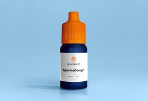
Slingshot’s SpectraComp® eGFP and mCherry Cell Mimics are ready-to-use multi-level fluorescent protein controls that mimic the autofluorescence and scatter profile of cells. These cell mimics use native eGFP and mCherry proteins that fluoresce in green and red fluorescent channels. Therefore, Slingshot’s SpectraComp® line of fluorescent proteins allows for accurate and consistent data interpretation without using biological samples for control purposes. Now you can accurately interpret the fluorescence signal in your sample, establish appropriate thresholds and validate functionality to ensure reliable and meaningful results.
Slingshot’s line of fluorescent protein cell mimics also offers a bright and stable fluorescence signal, providing a reliable reference for spectral unmixing and compensation. This allows for accurately determining the spectral overlap and spillover between fluorochromes in your high-parameter analyses.
Multiple workflow options offer greater flexibility, as SpectraComp® eGFP and SpectraComp® mCherry are provided in kits containing a negative population with three intensity levels (see below).
.png?width=1200&height=400&name=Introducing%20HyParComp%20(7).png)
Join Christian Nielsen, PhD of Odense University Hospital, Department of Clinical Immunology, and others in experiencing the value of Slingshot’s SpectraComp line of fluorescent cell mimics. Christian states, "it is just so great that you provide a separate tube with negative beads and that you have so low autofluorescence on your beads.”
Discover how to save time without compromising data accuracy with SpectraComp® eGFP and mCherry cell mimics.


.png?width=50&name=Sarah_Kotanchiyev%20(1).png)
